Ceevra for Thoracic Surgery
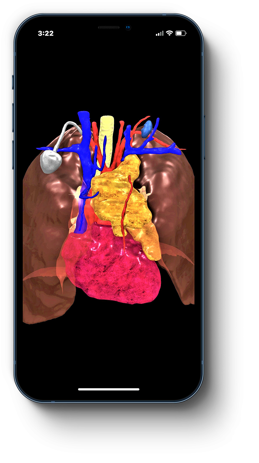
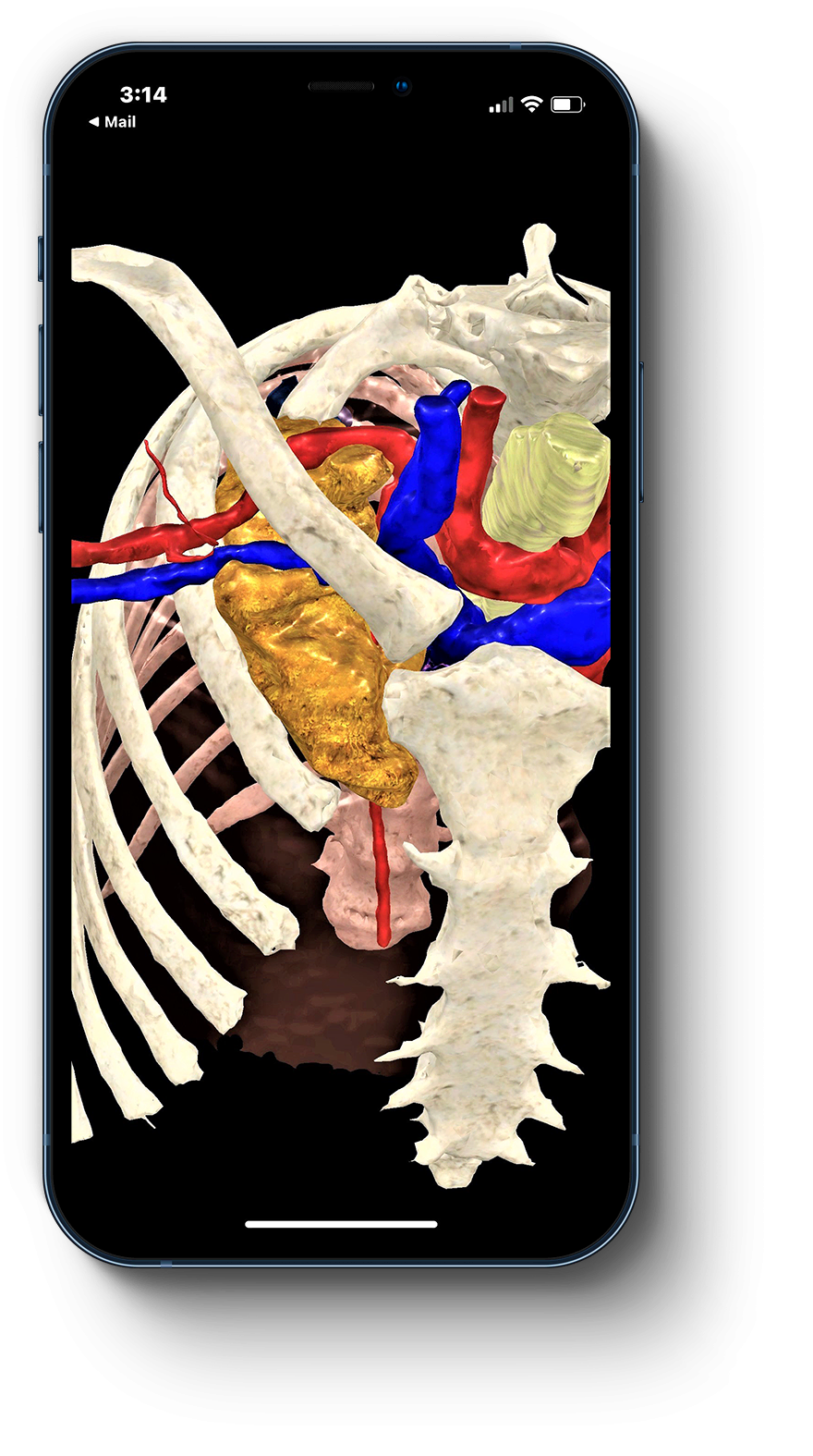
Ceevra 3D models are ideal for thoracic resections, including lung, mediastinal and chest wall lesions.
Ceevra in context: Thoracic Surgery
Thoracic surgeon Dr. Bernard Park details a sublobar resection aided by Ceevra
Sublobar Resection
For sublobar resections, we’ve developed novel, patented technology to help surgeons understand the segments involved and achieve the desired margins.
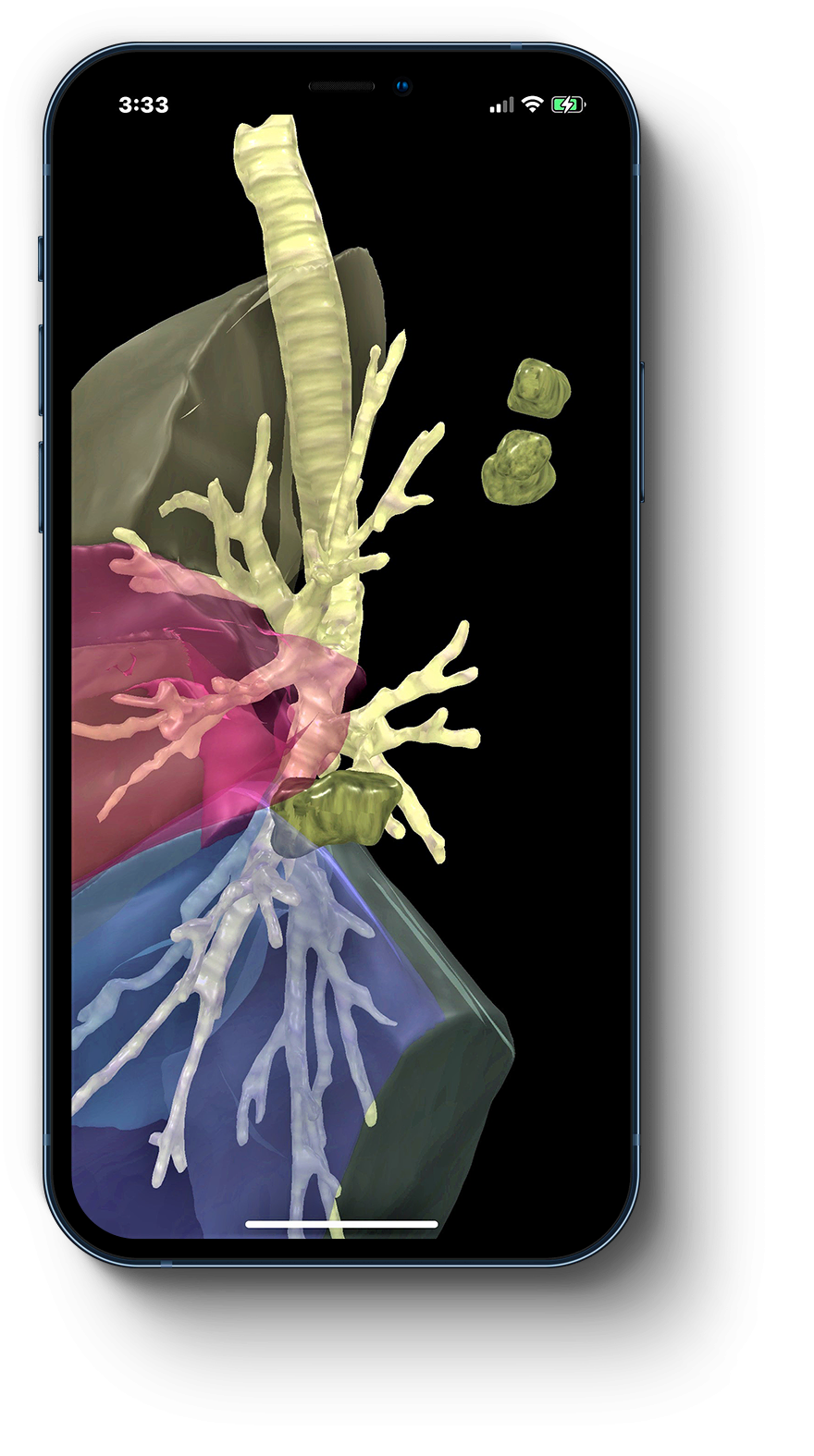
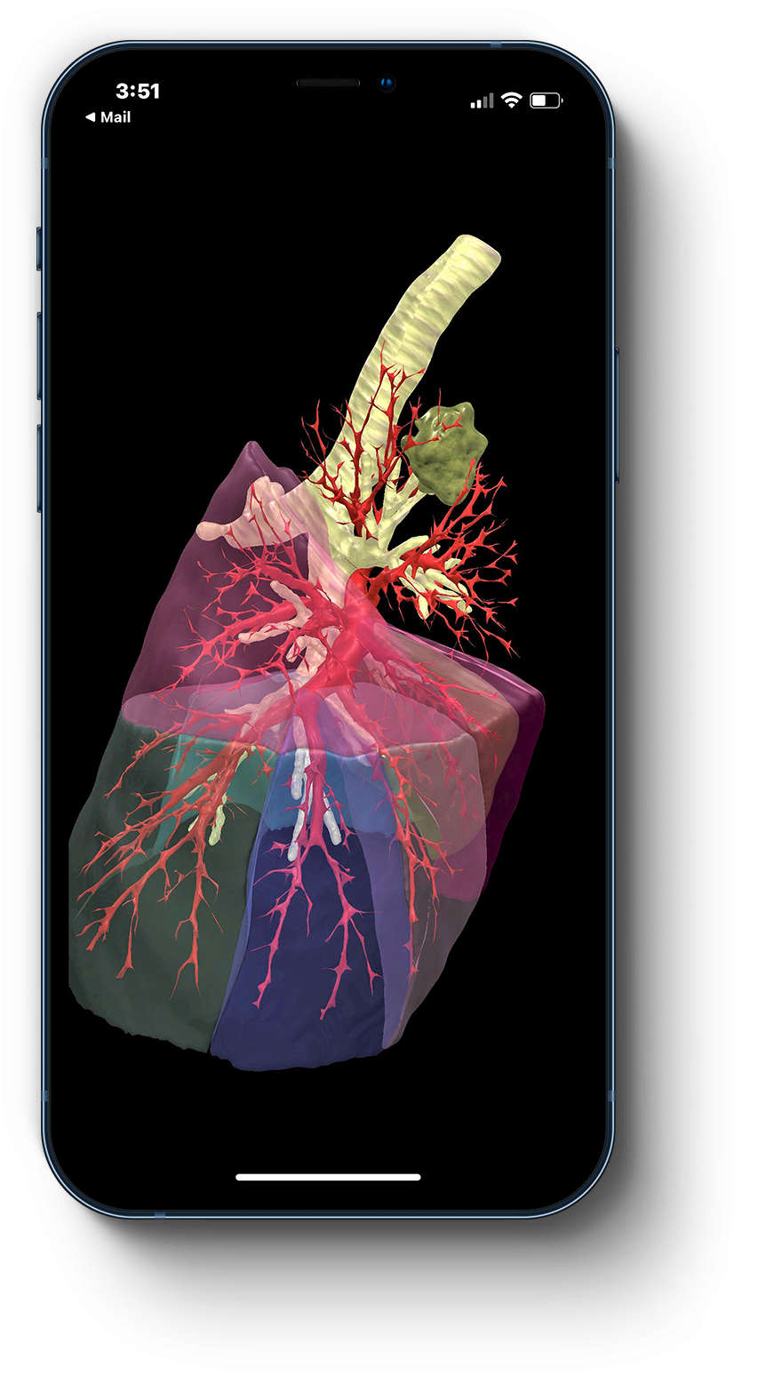
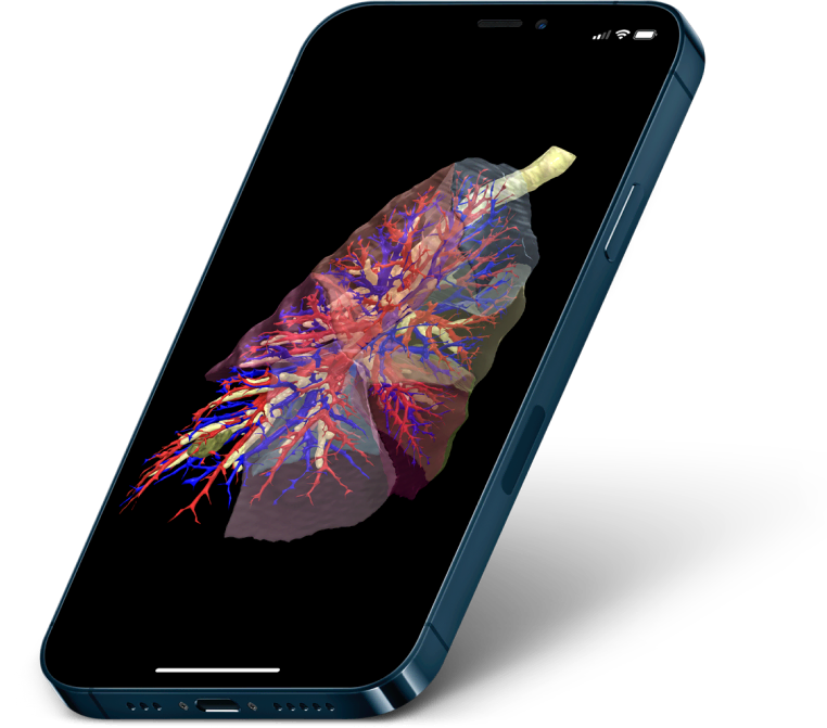
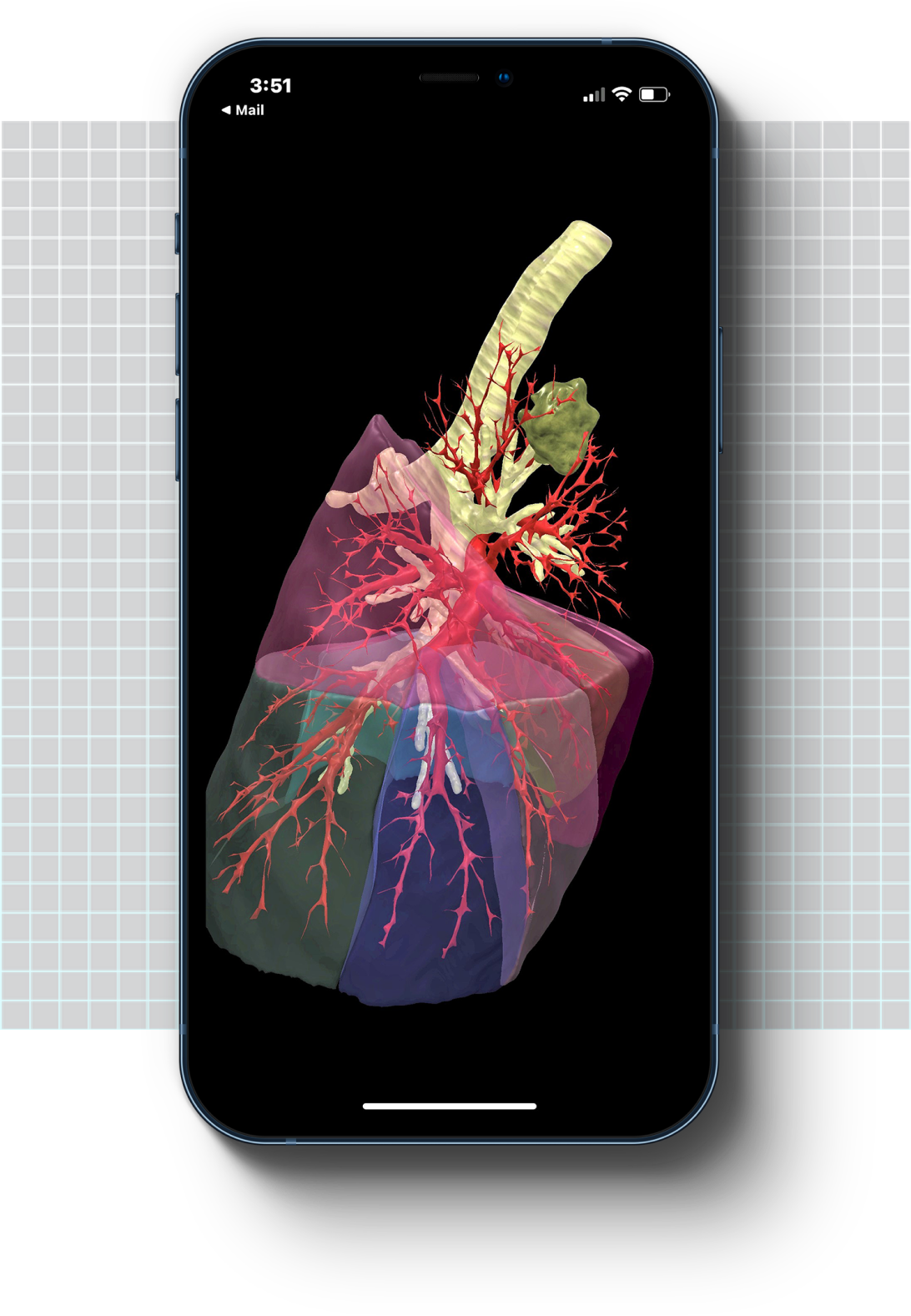
Lung 3D models down to the segmental level
Our software analyzes the patient’s pulmonary anatomy in the CT scan. It then delineates, displays and labels each individual lung segment as part of the 3D model.
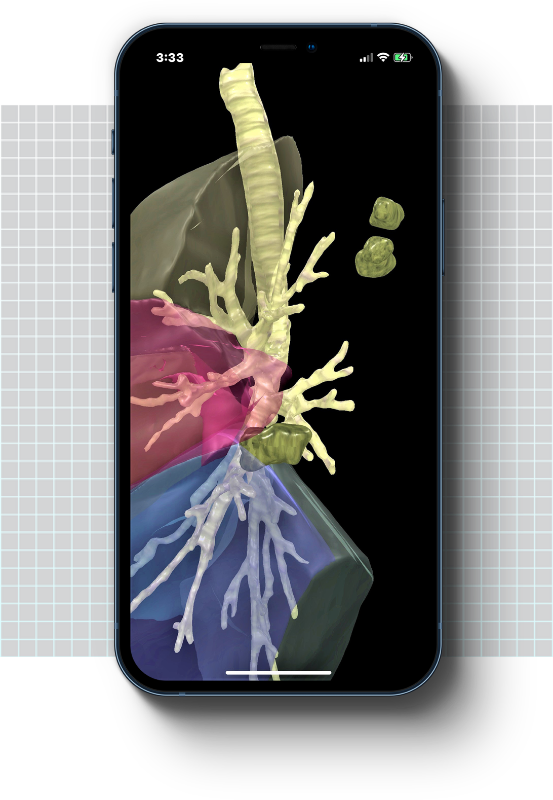
Customized View
Surgeons can selectively show/hide anatomical structures – including segments, veins and arteries – to clearly visualize the location of nodules relative to segmental boundaries.

“The Ceevra models show you extremely well where your margin’s going to be most at risk.”
Bernard J. Park, MD, FACS, FCCP
Thoracic Surgeon
Memorial Sloan Kettering Cancer Center
Supported thoracic SURGERY case types include: